How does reproductive acupuncture compares to the medication at improving ovulation?
One of the causes of anovulation is luteinised unruptured follicle syndrome (LUFS).
LUFS affects 5%-10% of healthy women and 25%-43% of women with infertility. This syndrome is more common among women diagnosed with endometriosis.
Most commonly LUFS is diagnosed during a series of ultrasound scans.
In women with LUFS, the dominant follicle will grow bigger than usual (up to 4 centimeters). The follicle will undergo the luteinisation process, but it will not rupture during the midcycle to release the mature egg. Progesterone levels will increase as if you have ovulated. Your endometrium will undergo premenstrual changes. In other words, you won’t be able to see any difference in your period. The menstrual flow will be no different from a healthy menstrual cycle. But, obviously, if the egg is not released, and if you have LUFS it is not, pregnancy cannot happen.
There is a clear explanation of why some women get LUFS. The main treatment for it is ovulation induction with hCG hormonal injection or Clomiphene. These treatments are a risk of hyperstimulation syndrome and other side-effects.
Does acupuncture offer a treatment alternative to anovulation?
A recent literature review on acupuncture’s effect on LUFS has been published in the Journal Of Acupuncture and Tuina Science. And if offers a safe and effective alternative. Read more
https://infertility-acupuncture.info/wp-content/uploads/2015/12/anovulation-LUFS-fertilisation-e1522116882445.jpg3991021Roberta Mekhttps://infertility-acupuncture.info/wp-content/uploads/2015/03/Infertility-acupuncture-logo-300x152.pngRoberta Mek2015-12-11 16:13:112021-09-29 13:58:35Reproductive acupuncture vs medication for anovulation
Acupuncture helps to normalise both ovarian volume and AMH in PCOS patients. These changes are beneficial for fertility.
Both acupuncture and exercise help to normalise overactive sympathetic nervous system in PCOS patients. Acupuncture, however, is much more effective. Furthermore, as shown by this study, exercise has no effect on AMH nor ovarian volume.
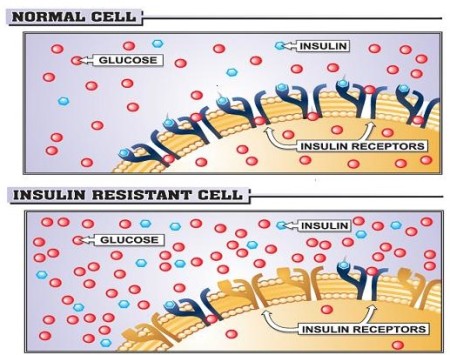
PCOS can improve with acupuncture treatment. More research looks into the mechanism of it. One of the lead researchers in PCOS and acupuncture Dr Elisabet Stener-Victorin from Sweden has long suspected that acupuncture may play a role in regulating blood glucose levels and helping with insulin resistance. Insulin resistance is one of the features of PCOS. This study put the theory to test.
The study revealed acupuncture increases whole-body glucose uptake during and after stimulation in women with polycystic ovary syndrome. See the abstract and a full text of the study below.
IVF Frozen Embryo Transfer (FET) patients benefit from Acupuncture. This helps us understand how Acupuncture benefits fertility.
Electro stimulation on acupuncture points leading to the IVF transfer improves the quality/receptivity of the uterine lining. A study published in the latest issue of peer-reviewed British Medical Journal (Acupuncture in Medicine) found the rates of embryo implantation, clinical pregnancy and live birth rates were higher in patients who received acupuncture leading to the transfer. They also found significant measurable changes in the endometrium (uterine lining):
Acupuncture improved the chances of triple-line pattern endometrial lining. It has been shown in studies that triple-line pattern is associated with good IVF outcome.
Endometrial perfusion (blood supply to the uterine lining) is an important factor in the process of implantation. The study found greater endometrial and subendometrial vascularisation following a series of acupuncture treatments leading to embryo transfer.
Acupuncture improved HOXA10 expression. Higher HOXA10 is associated with greater endometrial receptivity and good pregnancy outcomes. HOXA10 expression is lower in the uteri of women with hydrosalpinx, PCOS, and endometriosis.
How much acupuncture should you have to see those enhancements to your fertility? Women in this study had six acupuncture sessions per cycle for three menstrual cycles.
https://infertility-acupuncture.info/wp-content/uploads/2014/10/Acupuncture-improves-receptivity-of-endometrial-lining-in-IVF-Frozen-Embryo-Transfer-FET.jpg7681137Vitalis Skiauterishttps://infertility-acupuncture.info/wp-content/uploads/2015/03/Infertility-acupuncture-logo-300x152.pngVitalis Skiauteris2014-10-15 20:28:452021-11-16 17:13:03Acupuncture improves receptivity of endometrial lining in IVF Frozen Embryo Transfer
This study by Beijing Hospital of Obstetrics and Gynecology has shown acupuncture to be superior to clomophene on ovulation and pregnancy rates in treatment of polycystic ovary syndrome.
Other studies have shown that unlike Clomophene, acupuncture can have long term effect on PCOS by reducing the sympathetic nervous tonus. Apart from study methodology issues, another limitation of this study is that it only looked into the short term effect, but didn’t evaluate the effectiveness over a longer term. However, this is another study adding to the body of evidence. The study was published in the journal of Zhongguo Zhen jiu (Chinese Journal of Acupuncture & Moxibustion [2013, 33(11):961-964]).
Efficacy and safety evaluation of acupuncture combined with auricular acupoint therapy in the treatment of polycystic ovary syndrome
Department of Acupuncture and Physical Therapy, Beijing Obstetrics and Gynecology Hospital, Capital Medical University, Beijing 100026, China. geyongchun1981@sina.com
Zhongguo Zhen jiu = Chinese Acupuncture & Moxibustion [2013, 33(11):961-964]
Type: Journal Article, English Abstract (lang: chi)
OBJECTIVE: To compare the efficacy differences between acupuncture combined with auricular acupoint therapy and clomiphene oral administration in the treatment of polycystic ovary syndrome (PCOS).
METHODS: One hundred cases of PCOS were randomized into two groups, 50 cases in each one. Acupuncture combined with auricular acupuncture group (group A): acupuncture was applied at Guanyuan (CV 4), Zhongji (CV 3) and Zigong (EX-CA 1), once daily; auricular point sticking was applied at Spleen (pi, CO13), Endocrine (neifenmi, CO18), Uterus and Kidney (shen, CO10), the plaster was changed once a week. Clomiphene group (group B): oral clomiphene was prescribed at the 5th day of the menstrual, for 5 consecutive days, totally 3 menstrual cycles was needed. The ovulation induction, pregnancy and menstruation resuming of patients in the two ‘ , The totally effective rate was 90.00% (45/50) in group A, which was group were observed and compared.
RESULTS: The totally effective rate was 90.0% (45/50) in group A, which was superior to 86.0% (43/50) in group B (P<0.05); the ovulation rate and pregnancy rate were 68.0% (34/50)and 64. 0% (32/50) in group A, which were superior to that of group B (all P<0. 05); the menstruation resuming rate was 94.00 (47/50) in group A, which was superior to 88.00 (44/50) in group B (P<0.05). No adverse effect was observed in group A, while in group B, varying degrees of nausea, vomiting, headache and dermatitis were observed in 29 cases, ovarian hyperstimulation syndrome (OHSS) like polycystic ovary was observed in 14 cases under the B ultrasound.
CONCLUSION: Acupuncture combined with auricular acupoint therapy has a better effect than clomiphene in the treatment of PCOS without any adverse effects.
https://infertility-acupuncture.info/wp-content/uploads/2015/03/Infertility-acupuncture-logo-300x152.png00Vitalis Skiauterishttps://infertility-acupuncture.info/wp-content/uploads/2015/03/Infertility-acupuncture-logo-300x152.pngVitalis Skiauteris2014-02-22 11:16:312021-09-29 13:38:53Acupuncture more effective than clomophene in treatment of pcos
A truly thorough, well-referenced paper exploring the mechanism and effects of acupuncture on PCOS. Written by leading PCOS researchers from the Karolinska Institute in Sweden.
Polycystic Ovary Syndrome: Effect and Mechanisms of Acupuncture for Ovulation Induction
Julia Johansson and Elisabet Stener-Victorin
Abstract
Polycystic ovary syndrome (PCOS), the most common endocrine disorder among women of reproductive age, is characterized by the coexistence of hyperandrogenism, ovulatory dysfunction, and polycystic ovaries (PCO). PCOS also represents the largest part of female oligoovulatory infertility, and the management of ovulatory and menstrual dysfunction comprises a third of the high costs of PCOS treatment. Current pharmacological and surgical treatments for reproductive symptoms are effective, however, associated with negative side effects, such as cardiovascular complications and multiple pregnancies. For menstrual irregularities and ovulation induction in women with PCOS, acupuncture has indicated beneficial effects. This review will focus on the results from randomized controlled acupuncture trials for the regulation of menstrual dysfunction and for inducing ovulation in women with PCOS although there are uncontrolled trials with nonetheless interesting results. Animal experimental studies will be further discussed when they can provide a more mechanistic explanatory view. Read more
https://infertility-acupuncture.info/wp-content/uploads/2015/03/Infertility-acupuncture-logo-300x152.png00Vitalis Skiauterishttps://infertility-acupuncture.info/wp-content/uploads/2015/03/Infertility-acupuncture-logo-300x152.pngVitalis Skiauteris2013-10-27 14:01:172021-09-29 13:39:22PCOS Acupuncture: the most comprehensive paper to date?
The short answer is yes, acupuncture can improve embryo quality. Now let’s have a look into study details.
Dr Rashidi and his team explored if acupuncture can improve pregnancy rates for PCOS patients undergoing IVF and ICSI. As a result, they found that acupuncture can improve embryo quality.
Acupuncture showed considerable advantages over the metformin for obese PCOS patients. Acupuncture was shown to be more effective then metformin at improving menstrual freqency, reducing body mass index and waist to hip ratio. It also had fewer side-effects.
Effectiveness of Abdominal Acupuncture for Patients with Obesity-Type Polycystic Ovary Syndrome: A Randomized Controlled Trial
To cite this article:
Yan-Hua Zheng, Xin-Hua Wang, Mao-Hua Lai, Hong Yao, Hua Liu, and Hong-Xia Ma. The Journal of Alternative and Complementary Medicine. -Not available-, ahead of print. doi:10.1089/acm.2012.0429.
https://infertility-acupuncture.info/wp-content/uploads/2015/03/Infertility-acupuncture-logo-300x152.png00Vitalis Skiauterishttps://infertility-acupuncture.info/wp-content/uploads/2015/03/Infertility-acupuncture-logo-300x152.pngVitalis Skiauteris2013-05-24 17:46:572021-09-29 13:39:34Acupuncture superior to Metformin for PCOS
Electrical and manual acupuncture stimulation affects estrous cyclicity and neuroendocrine function in a DHT-induced rat polycystic ovary syndrome model
Yi Feng, Julia Johansson, Ruijin Shao, Louise Mannerås Holm, Håkan Billig, Elisabet Stener-Victorin
Abstract
Both low-frequency electro-acupuncture (EA) and manual acupuncture improve menstrual frequency and decrease circulating androgens in women with polycystic ovary syndrome (PCOS). We sought to determine whether low-frequency EA is more effective than manual stimulation in regulating disturbed estrous cyclicity in rats with PCOS induced by 5?-dihydrotestosterone (DHT). To identify the central mechanisms of the effects of stimulation, we assessed hypothalamic mRNA expression of molecules that regulate reproductive and neuroendocrine function.
From age 70 days, rats received 2-Hz EA or manual stimulation of the needles five times/week for 4–5 weeks; untreated rats served as controls. Specific hypothalamic nuclei were obtained by laser microdissection, and mRNA expression was measured with TaqMan low-density arrays. Untreated rats were acyclic. During the last 2 weeks of treatment, seven of eight (88%) rats in the EA group had epithelial keratinocytes, demonstrating estrous cycle change (p= 0.034 vs. controls). In the manual group, five of eight (62%) rats had estrous cycle changes (ns vs. controls). mRNA expression of the opioid receptors Oprk1 and Oprm1 in the hypothalamic arcuate nucleus was lower in the EA group than in untreated controls. mRNA expression of the steroid hormone receptors Esr2, Pgr, and Kiss1r was lower in the manual group than in the controls.
In rats with DHT-induced PCOS, low-frequency EA restored disturbed estrous cyclicity but did not differ from manual stimulation group, although electrical stimulation lowered serum testosterone in responders, those with restored estrus cyclicity, and differed from both controls and the manual stimulation group. Thus, EA cannot in all aspects be considered superior to manual stimulation.
The effects of low-frequency EA may be mediated by central opioid receptors, while manual stimulation may involve regulation of steroid hormone/peptide receptors.
https://infertility-acupuncture.info/wp-content/uploads/2015/03/Infertility-acupuncture-logo-300x152.png00Vitalis Skiauterishttps://infertility-acupuncture.info/wp-content/uploads/2015/03/Infertility-acupuncture-logo-300x152.pngVitalis Skiauteris2012-02-08 15:58:582021-09-26 12:32:03Acupuncture improves menstrual frequency and decreases androgens in women with polycystic ovary syndrome (PCOS)
Title: Effect of electro-acupuncture on ovarian expression of ? (1)- and ? (2)-adrenoceptors, and p75 neurotrophin receptors in rats with steroid-induced polycystic ovaries
Author: Manni Luigi ; Lundeberg Thomas ; Holmäng Agneta ; Aloe Luigi ; Stener-Victorin Elisabet
Abstract:
Abstract
Background
Estradiol valerate (EV)-induced polycystic ovaries (PCO) in rats is associated with an increase in ovarian sympathetic outflow. Low-frequency (2 Hz) electro-acupuncture (EA) has been shown to modulate sympathetic markers as well as ovarian blood flow as a reflex response via the ovarian sympathetic nerves, in rats with EV-induced PCO.
Methods
In the present study, we further tested the hypothesis that repeated 2 Hz EA treatments modulate ovarian sympathetic outflow in rats with PCO, induced by a single i.m. injection of EV, by investigating the mRNA expression, the amount and distribution of proteins of ?1a-, ?1b-, ?1d-, and ?2-adrenoceptors (ARs), as well as the low-affinity neurotrophin receptor (p75NTR).
Results
It was found that EV injection results in significantly higher mRNA expression of ovarian ?1b- and ?1d-AR in PCO rats compared to control rats. The p75NTR and ?2-ARs mRNA expression were unchanged in the PCO ovary. Low-frequency EA resulted in a significantly lower expression of ?2-ARs mRNA expression in PCO rats. The p75NTR mRNA was unaffected in both PCO and control rats. PCO ovaries displayed significantly higher amount of protein of ?1a-, ?1b- and ?1d-ARs, and of p75NTR, compared to control rats, that were all counteracted by repeated low-frequency EA treatments, except for ?1b-AR.
Conclusion
The present study shows that EA normalizes most of the EV-induced changes in ovarian ARs. Furthermore, EA was able to prevent the EV-induced up regulation of p75NTR, probably by normalizing the sympathetic ovarian response to NGF action. Our data indicate a possible role of EA in the regulation of ovarian responsiveness to sympathetic inputs and depict a possible complementary therapeutic approach to overcoming sympathetic-related anovulation in women with PCOS.
Journal: Reproductive Biology and Endocrinology
Issn: 14777827
https://infertility-acupuncture.info/wp-content/uploads/2015/03/Infertility-acupuncture-logo-300x152.png00Vitalis Skiauterishttps://infertility-acupuncture.info/wp-content/uploads/2015/03/Infertility-acupuncture-logo-300x152.pngVitalis Skiauteris2011-08-13 16:43:562021-09-24 08:58:22Acupuncture normalizes most of the Estradiol Valerate induced changes in ovarian adresnoceptors
Title: Effect of electro-acupuncture stimulation of different frequencies and intensities on ovarian blood flow in anaesthetized rats with steroid-induced polycystic ovaries
Author: Stener-Victorin Elisabet ; Kobayashi Rie ; Watanabe Orie ; Lundeberg Thomas ; Kurosawa Mieko
Abstract:
Abstract
Background
Maintenance of ovarian blood flow (OBF) is suggested to be important for regular ovulation in women with polycystic ovaries (PCO). Read more
Current evidence of acupuncture on polycystic ovarian syndrome.
Lim CE, Wong WS.
Faculty of Medicine, South Western Sydney Clinical School, University of New South Wales, Sydney, Australia. celim@unswalumni.com Gynecol Endocrinol. 2010 Jun;26(6):473-8.
Abstract
OBJECTIVE: This paper aims to provide a literature review on evaluating the efficacy of acupuncture therapy in the treatment of polycystic ovarian syndrome (PCOS) by reviewing clinical trials; randomised and non-randomised and observational studies on PCOS. The paper will also determine the possible mechanism of acupuncture treatment in PCOS, limitations of recruited studies and suggest further improvements in future studies. Read more
https://infertility-acupuncture.info/wp-content/uploads/2015/03/Infertility-acupuncture-logo-300x152.png00Vitalis Skiauterishttps://infertility-acupuncture.info/wp-content/uploads/2015/03/Infertility-acupuncture-logo-300x152.pngVitalis Skiauteris2011-02-19 15:28:062021-09-26 12:31:31Acupuncture is a safe and effective treatment for PCOS (Polycystic Ovary Syndrome)
Impact of electro-acupuncture and physical exercise on hyperandrogenism and oligo/amenorrhea in women with polycystic ovary syndrome: a randomized controlled trial
Elizabeth Jedel1, Fernand Labrie2, Anders Odén3, Göran Holm4, Lars Nilsson5, Per Olof Janson5, Anna-Karin Lind5, Claes Ohlsson6, and Elisabet Stener-Victorin7,8 Read more
https://infertility-acupuncture.info/wp-content/uploads/2015/03/Infertility-acupuncture-logo-300x152.png00Vitalis Skiauterishttps://infertility-acupuncture.info/wp-content/uploads/2015/03/Infertility-acupuncture-logo-300x152.pngVitalis Skiauteris2011-02-10 18:13:372021-09-29 13:58:11Acupuncture improves hyperandrogenism and menstrual frequency in women with PCOS
Low-frequency Electro-Acupuncture and Physical Exercise Decrease High Muscle Sympathetic Nerve Activity in Polycystic Ovary Syndrome
Elisabet Stener-Victorin1*, Elizabeth Jedel2, Per Olof Janson, and Yrsa Bergmann Sverrisdottir3
1 Institution of Neuroscience and Physiology
2 Osher Center for Integrative Medicine
3 inst. neuroscience and physiology
* To whom correspondence should be addressed. E-mail: elisabet.stener-victorin@neuro.gu.se.
Context: We have recently shown that polycystic ovary syndrome (PCOS) is associated with high muscle sympathetic nerve activity. Animal studies support the concept that low-frequency electro-acupuncture (EA) and physical exercise, via stimulation of ergoreceptors and somatic afferents in the muscles, may modulate the activity of the sympathetic nervous system. Objective: The aim of the present study was to investigate the effect of these interventions on sympathetic nerve activity in women with PCOS. Design: Randomized controlled trial. Setting: Sahlgrenska University Hospital, Gothenburg, Sweden. Outcome Measures and Subjects: Twenty women with PCOS were randomly allocated to one of three groups; low-frequency EA (n=9), physical exercise (n=5) or to an untreated control (n=6) group during 16 weeks. Direct recordings of multiunit efferent postganglionic muscle sympathetic nerve activity (MSNA) in a muscle fascicle of the peroneal nerve before and following 16 weeks of treatment. Biometric, hemodynamic, endocrine and metabolic parameters were measured. Results: Low-frequency EA (P = 0.036) and physical exercise (P = 0.030) decreased MSNA burst frequency compared to the untreated control group. Low-frequency EA group reduced sagittal diameter (P = 0.001), while physical exercise group reduced body weight (P = 0.004) and body mass index (BMI) (P = 0.004) as compared to the untreated control group. Sagittal diameter was related to MSNA burst frequency (Rs = 0.58, P < 0.005) in the EA group. No correlation was found for BMI and MSNA in the exercise group. There were no differences between the groups in hemodynamic, endocrine and metabolic variables. Conclusions: For the first time we demonstrate that low-frequency EA and physical exercise lowers high sympathetic nerve activity in women with PCOS. Thus, treatment with low-frequency EA or physical exercise with the aim to reduce MSNA may be of importance for women with PCOS.
Am J Physiol Regul Integr Comp Physiol (June 3, 2009). doi:10.1152/ajpregu.00197.2009
Acupuncture and exercise restore adipose tissue expression of sympathetic markers and improve ovarian morphology in rats with dihydrotestosterone-induced PCOS
Louise Mannerås,1 Stefan Cajander,2 Malin Lönn,3 and Elisabet Stener-Victorin1
1Institute of Neuroscience and Physiology, Department of Physiology, Sahlgrenska Academy, University of Gothenburg, Gothenburg, Sweden; 2 Department of Pathology and Cytology, Sunderby County Hospital, Luleå, Sweden; and 3 Institute of Medicine, Wallenberg Laboratory, Sahlgrenska Academy, University of Gothenburg, Gothenburg, Sweden
Submitted 22 November 2008 ; accepted in final form 15 January 2009
Altered activity of the sympathetic nervous system, which innervates adipose and ovarian tissue, may play a role in polycystic ovary syndrome (PCOS). We hypothesize that electro-acupuncture (EA) and physical exercise reduce sympathetic activity by stimulating ergoreceptors and somatic afferent pathways in muscles. Here we investigated the effects of low-frequency EA and physical exercise on mRNA expression of sympathetic markers in adipose tissue and on ovarian morphology in female rats that received dihydrotestosterone (DHT) continuously, starting before puberty, to induce PCOS. At age 11 wk, rats with DHT-induced PCOS were randomly divided into three groups: PCOS, PCOS plus EA, and PCOS plus exercise. The latter two groups received 2-Hz EA (evoking muscle twitches) three times/week or had free access to a running wheel for 4–5 wk. In mesenteric adipose tissue, expression of ?3-adrenergic receptor (ADRB3), nerve growth factor (NGF), and neuropeptide Y (NPY) mRNA was higher in untreated PCOS rats than in controls. Low-frequency EA and exercise downregulated mRNA expression of NGF and NPY, and EA also downregulated expression of ADRB3, compared with untreated rats with DHT-induced PCOS. EA and exercise improved ovarian morphology, as reflected in a higher proportion of healthy antral follicles and a thinner theca interna cell layer than in untreated PCOS rats. These findings support the theory that increased sympathetic activity contributes to the development and maintenance of PCOS and that the effects of EA and exercise may be mediated by modulation of sympathetic outflow to the adipose tissue and ovaries.
The Role of Acupuncture in the Management of Subfertility
Ng E H et al Fertil Steril. 2008 Jul;90(1):1-13.
Fertility and Sterility
Abstract
OBJECTIVE: To review systematically the use of acupuncture in the management of subfertility.
DESIGN: A computer search was performed via several English and Chinese databases to identify journals relevant to the subject.
RESULT(S): The positive effect of acupuncture in the treatment of subfertility may be related to the central sympathetic inhibition by the endorphin system, the change in uterine blood flow and motility, and stress reduction. Acupuncture may help restore ovulation in patients with polycystic ovary syndrome, although there are not enough randomized studies to validate this.
There is also no sufficient evidence supporting the role of acupuncture in male subfertility, as most of the studies are uncontrolled case reports or case series in which the sample sizes were small. Despite these deficiencies, acupuncture can be considered as an effective alternative for pain relief during oocyte retrieval in patients who cannot tolerate side effects of conscious sedation.
The pregnancy rate of IVF treatment is significantly increased, especially when acupuncture is administered on the day of embryo transfer.
CONCLUSION(S): Although acupuncture has gained increasing popularity in the management of subfertility, its effectiveness has remained controversial.
https://infertility-acupuncture.info/wp-content/uploads/2015/03/Infertility-acupuncture-logo-300x152.png00Vitalis Skiauterishttps://infertility-acupuncture.info/wp-content/uploads/2015/03/Infertility-acupuncture-logo-300x152.pngVitalis Skiauteris2008-09-24 14:31:362021-09-24 08:58:33Acupuncture for male and female infertility – systematic review
Institute of Neuroscience and Physiology, Department of Physiology, Sahlgrenska Academy, Göteborg University, Göteborg, Sweden. elisabet.stener-victorin@neuro.gu.se
Abstract
This review describes the aetiology and pathogenesis of polycystic ovary syndrome (PCOS) and evaluates the use of acupuncture to prevent and reduce symptoms related with PCOS. PCOS is the most common female endocrine disorder and it is strongly associated with hyperandrogenism, ovulatory dysfunction and obesity. PCOS increases the risk for metabolic disturbances such as hyperinsulinaemia and insulin resistance, which can lead to type 2 diabetes, hypertension and an increased likelihood of developing cardiovascular risk factors and impaired mental health later in life. Despite extensive research, little is known about the aetiology of PCOS. The syndrome is associated with peripheral and central factors that influence sympathetic nerve activity. Thus, the sympathetic nervous system may be an important factor in the development and maintenance of PCOS. Many women with PCOS require prolonged treatment. Current pharmacological approaches are effective but have adverse effects. Therefore, nonpharmacological treatment strategies need to be evaluated. Clearly, acupuncture can affect PCOS via modulation of endogenous regulatory systems, including the sympathetic nervous system, the endocrine and the neuroendocrine system. Experimental observations in rat models of steroid-induced polycystic ovaries and clinical data from studies in women with PCOS suggest that acupuncture exert long-lasting beneficial effects on metabolic and endocrine systems and ovulation.
https://infertility-acupuncture.info/wp-content/uploads/2015/03/Infertility-acupuncture-logo-300x152.png00Vitalis Skiauterishttps://infertility-acupuncture.info/wp-content/uploads/2015/03/Infertility-acupuncture-logo-300x152.pngVitalis Skiauteris2008-03-21 21:45:292021-09-24 08:59:38Acupuncture in polycystic ovary syndrome: current experimental and clinical evidence.
Effects of acupuncture on rates of pregnancy and live birth among women undergoing in vitro fertilisation: systematic review and meta-analysis
Eric Manheimer, research associate1, Grant Zhang, assistant professor1, Laurence Udoff, assistant professor2, Aviad Haramati, professor3, Patricia Langenberg, professor and vice-chair4, Brian M Berman, professor1, Lex M Bouter, professor and vice chancellor (rector magnificus)5
1 Center for Integrative Medicine, University of Maryland School of Medicine, 2200 Kernan Drive, Kernan Hospital Mansion, Baltimore, MD 21207, USA, 2 Department of Obstetrics, Gynecology and Reproductive Services, University of Maryland School of Medicine, 3 Department of Physiology and Biophysics and Medicine, Georgetown University School of Medicine, Washington, DC, 4 Department of Epidemiology and Preventive Medicine, University of Maryland School of Medicine, 5 VU University Amsterdam De Boelelaan 1105, 1081 HV Amsterdam, the Netherlands
Correspondence to: E Manheimer emanheimer@compmed.umm.edu
Objective To evaluate whether acupuncture improves rates of pregnancy and live birth when used as an adjuvant treatment to embryo transfer in women undergoing in vitro fertilisation.
Design Systematic review and meta-analysis.
Data sources Medline, Cochrane Central, Embase, Chinese Biomedical Database, hand searched abstracts, and reference lists.
Review methods Eligible studies were randomised controlled trials that compared needle acupuncture administered within one day of embryo transfer with sham acupuncture or no adjuvant treatment, with reported outcomes of at least one of clinical pregnancy, ongoing pregnancy, or live birth. Two reviewers independently agreed on eligibility; assessed methodological quality; and extracted outcome data. For all trials, investigators contributed additional data not included in the original publication (such as live births). Meta-analyses included all randomised patients.
Data synthesis Seven trials with 1366 women undergoing in vitro fertilisation were included in the meta-analyses. There was little clinical heterogeneity. Trials with sham acupuncture and no adjuvant treatment as controls were pooled for the primary analysis. Complementing the embryo transfer process with acupuncture was associated with significant and clinically relevant improvements in clinical pregnancy (odds ratio 1.65, 95% confidence interval 1.27 to 2.14; number needed to treat (NNT) 10 (7 to 17); seven trials), ongoing pregnancy (1.87, 1.40 to 2.49; NNT 9 (6 to 15); five trials), and live birth (1.91, 1.39 to 2.64; NNT 9 (6 to 17); four trials). Because we were unable to obtain outcome data on live births for three of the included trials, the pooled odds ratio for clinical pregnancy more accurately represents the true combined effect from these trials rather than the odds ratio for live birth. The results were robust to sensitivity analyses on study validity variables. A prespecified subgroup analysis restricted to the three trials with the higher rates of clinical pregnancy in the control group, however, suggested a smaller non-significant benefit of acupuncture (odds ratio 1.24, 0.86 to 1.77).
Conclusions Current preliminary evidence suggests that acupuncture given with embryo transfer improves rates of pregnancy and live birth among women undergoing in vitro fertilisation.
https://infertility-acupuncture.info/wp-content/uploads/2015/03/Infertility-acupuncture-logo-300x152.png00Vitalis Skiauterishttps://infertility-acupuncture.info/wp-content/uploads/2015/03/Infertility-acupuncture-logo-300x152.pngVitalis Skiauteris2008-02-10 10:08:432021-09-24 08:59:16Systematic review: Acupuncture and IVF: Acupuncture improves rates of pregnancy and live birth
Effect of electro-acupuncture stimulation of different frequencies and intensities on ovarian blood flow in anaesthetized rats with steroid-induced polycystic ovaries
Maintenance of ovarian blood flow (OBF) is suggested to be important for regular ovulation in women with polycystic ovaries (PCO). The purpose of the present study was to investigate whether electro-acupuncture (EA) of different frequencies and intensities can improve the OBF of anaesthetized rat in the animal model of PCO.
Methods
PCO was experimentally induced by a single intramuscular (i.m.) injection of estradiol valerate (EV) in rats. Control rats were given i.m. injection of oil. The involvement of the two ovarian sympathetic nerves; superior ovarian nerve (SON) and plexus ovarian nerve (OPN), in OBF responses was elucidated by severance of SON and OPN in both control and PCO rats. How systemic circulatory changes affect OBF was evaluated by continuous recording of the blood pressure. OBF was measured on the surface of the ovary-using laser Doppler flowmetry. Acupuncture needles were inserted bilaterally into the abdominal and hind limb muscles and connected to an electrical stimulator. Two frequencies – 2 Hz (low) and 80 Hz (high) – with three different intensities – 1.5, 3, and 6 mA – were applied for 35 s.
Results
Low-frequency EA at intensities of 3 and 6 mA elicited significant increases in OBF in the Control group compared to baseline. In the PCO group the increases in OBF were significant only when stimulating with low-frequency EA at 6 mA. After severance of the ovarian sympathetic nerves, the increased response of OBF that had been induced by low-frequency EA in both the Control and PCO group was abolished, indicating that the OBF response is mediated via the ovarian sympathetic nerves. High-frequency EA at 6 mA significantly decreased OBF and mean arterial blood pressure (MAP) in the Control group compared to baseline. In the PCO group, the same stimulation produced similar decreases in MAP, but not in OBF.
Conclusion
Low-frequency EA stimulation with a strong intensity (6 mA) increases OBF in rats with steroid-induced PCO whereas less strong intensity (3 mA) induces similar changes in control rats. Severance of the ovarian sympathetic nerves, abolish this OBF increase in both study groups, which suggests that the responses of OBF to EA are mediated via the ovarian sympathetic nerves. Read more
Clinical studies on the mechanism for acupuncture stimulation of ovulation
Mo X; Li D; Pu Y; Xi G; Le X; Fu Z Zhejiang College of Traditional Chinese Medicine, Hangzhou.
Ovulatory dysfunction is commonly seen in gynecology clinic. It may cause infertility, amenia, functional uterine bleeding and a variety of complications. This research according to TCM theory records treating with acupuncture 34 patients suffering from ovulatory dysfunction. Changes in clinical symptoms and some relative targets are reported, plus findings in animal experiments. The theory concerning the generative and physiologic axis of women, this research involved the following points; Ganshu (UB 18), Shenshu (UB 23), Guanyuan (Ren 4), Zhongji (Ren 3), and Sanyinjiao (Sp 6). The reinforcement and reduction of acupuncture enables it to strengthen liver and kidney. Through the Chong and Ren channels it nourishes uterus to adjust the patient’s axis function and recover ovulation. Treated on an average of 30 times, the patients’ symptoms improved to varying degrees. The marked effective rate was 35.29%, the total effective rate being 82.35%. BBT, VS, CMS, and B ultrasonic picture all improved to some degree. The results also showed that acupuncture may adjust FSH, LH, and E2 in two directions and raise the progesterone level, bringing them to normal. The animal experiments confirmed this result. Results showed that acupuncture may adjust endocrine function of the generative and physiologic axis of women, thus stimulating ovulation. The results of this research will provide some scientific basis for treating and further studying this disorder.


 This study by Beijing Hospital of Obstetrics and Gynecology has shown acupuncture to be superior to clomophene on ovulation and pregnancy rates in treatment of polycystic ovary syndrome.
 Acupuncture showed considerable advantages over the metformin for obese PCOS patients. Acupuncture was shown to be more effective then metformin at improving menstrual freqency, reducing body mass index and waist to hip ratio. It also had fewer side-effects.


